SPIRIT Instructions For Use
|
Gold Standard Phantoms |
Document ID: PD-3011-0007 |
| Revision | Summary | Date |
|---|---|---|
| 1.0 | First issue. | 18 Nov 2025 |
Foreword
These instructions for use contain all the information necessary to operate the SPIRIT phantom in accordance with its specifications. This information includes explanations of the functions of the controls, displays and signals, the sequence of operation, and connection and disconnection of the parts and accessories you can remove.
You must regard these instructions as a part of the equipment. It is important that these instructions are read thoroughly.
Disclaimer
Gold Standard Phantoms considers itself responsible for the effects on safety, reliability, and performance of the equipment only if:
Assembly operations, re-adjustments, modification or repairs are carried out by persons authorised by ourselves, and
The electrical installation of the room where the device is used meets the requirements of the standards in force, and
The device is used in accordance with this instruction manual.
Glossary and Abbreviations
Term | Description |
|---|---|
MRI | Magnetic Resonance Imaging |
SPIRIT | Spherical multiParameter Imaging Reference for Instrument Testing |
MR | Magnetic Resonance |
Diffusion | Random movements of a molecule in an isotropic solution without thermal gradients. In MR typically this refers to the self-diffusion of water molecules. |
Relaxometry | Measurements of the MR relaxation times |
T1 | Longitudinal (spin-lattice) relaxation time constant |
T2 | Transverse (spin-spin) relaxation time constant |
D | Self-diffusivity of water |
HDPE | High Density Polyethylene |
PETG | Polyethylene terephthalate glycol |
EVA | Ethylene Vinyl Acetate |
FKM | Fluorelastomer |
SDS | Safety Data Sheet |
EG | Ethylene Glycol |
Introduction
About this Instruction Manual
The purpose of this instruction manual is to promote the safe use of a SPIRIT phantom during its expected service life.
It describes the use of the device as intended by the manufacturer.
Identification
Product Name: SPIRIT
Manufacturer and Contact Information
Company Identification | Gold Standard Phantoms Limited 1A Parkway Rise Sheffield South Yorkshire S9 4WQ United Kingdom |
Telephone | +44 (0)114 3271540 |
Email (competent person) | |
Languages spoken | English |
EU Authorised Representative | AR Experts B.V. |
EU Authorised Representative Address | AR Experts B.V. Boeingavenue 201-219 1119 PD Schiphol-Rijk The Netherlands |
EU Authorised Representative Email Address |
Intended Use
The SPIRIT phantom is designed for Quality Assurance measurements of Magnetic Resonance Imaging (MRI). Phantoms (also known as Quality Assurance Devices) are used in lieu of a human being inside an MRI scanner to assess its performance. The user should refer to the MRI device’s instructions for use to set up the correct pulse sequence for a given outcome. Equivalent acquisition techniques need to be used on the phantom as in the humans / patients.
Intended User
The SPIRIT phantom is intended for use by qualified MRI system operating personnel. The phantom must be used by competent personnel who have the applicable education and experience. This includes but is not limited to:
Radiographer/MRI Technologist
Clinical Scientist/Hospital Physicist
MRI Physicist
Radiologist
Student/Researcher
Other trained Medical Professional.
As for any phantom, this device must be used by a skilled person who has the applicable education and experience to enable them to perceive risks and to avoid hazards which operation or maintenance of the device can create.
Warnings and Cautions Used
Essential safety descriptions are provided in the instruction manual in the form of either a Warning or a Caution, as described below:
A WARNING is given when the personal safety of the participant or a user can be affected. Disregarding this advice can cause an injury.
A CAUTION is given when special instructions must be followed. Disregarding this advice can cause damage to the device or other equipment.
All users must familiarise themselves with all the warnings and cautions contained in the instructions before using this device.
Device Description
About the SPIRIT
SPIRIT
The SPIRIT is a 191 mm spherical phantom designed for use in an MRI scanner. The SPIRIT holds a total of sixteen 4ml Nalgene vials and four 10 mm Polypropylene vials. These vials contain a set of solutions with known reference quantities. It additionally contains an array of physical features which can be used for assessing the performance of MRI scanners. Further, the Phantom contains a pair of ethylene glycol filled NMR tubes which can be used to determine the temperature via MR thermometry.
It addresses the need for a standardised neuroimaging test object for multisite validation of advanced MRI techniques.
Applications include:
Quantitative T1/T2 relaxometry
Quantitative Diffusion imaging
Mapping of geometric distortions due to gradient field non-linearities
Measurement of effective slice thickness
Assessment of effective resolution
Materials
Component | Materials Used |
|---|---|
Main shell and Sample Holder | Polycarbonate |
Vials | Polypropylene and HDPE |
Cap | Polypropylene, Nitrile rubber, Nylon |
Fiducal grid | Accura ClearVue |
Phantom liquid | Distilled Water, Manganese (II) Chloride, Surfactant (C8-18 Ethoxylated Propoxylated), and preservative (Methylisothiazolinone and Chloromethylisothiazolinone) |
Vial Contents | PVP, Nickel (II) Chloride |
NMR tube material | Borosilicate glass with EVA cap |
NMR tube contents | Ethylene glycol |
Phantom Holders | PETG, Nylon |
WARNING: The phantom liquid is a mild irritant and may be skin sensitising. In the event of a leak or spill of the phantom liquid wear protective waterproof gloves when cleaning any spillages and dispose according to local regulations. See the safety data sheet for more information.
About the Vials
The phantom contains a set of Prefilled Vials. These plastic vials are filled with reference materials for Quality Assurance measurements in Magnetic Resonance Imaging. The vials contained in the SPIRIT come in two sizes:

Vial ID | Type | Product Code | Description | Vial Size | SI Traceable Characterisation Available |
A | Relaxometry | MNCL-0320 | 0.320mM Aqueous MnCl2 | 10 ml | T1, T2 |
B | Relaxometry | MNCL-0159 | 0.159mM Aqueous MnCl2 | 10 ml | T1, T2 |
C | Relaxometry | MNCL-0110 | 0.110mM Aqueous MnCl2 | 10 ml | T1, T2 |
D | Relaxometry | MNCL-0039 | 0.039mM Aqueous MnCl2 | 10 ml | T1, T2 |
E | Diffusion | PVP-0050 | 5% Aqueous PVP NiCl2 | 4 ml | T1, T2, D |
F | Diffusion | PVP-0100 | 10% Aqueous PVP NiCl2 | 4 ml | T1, T2, D |
G | Diffusion | PVP-0150 | 15% Aqueous PVP NiCl2 | 4 ml | T1, T2, D |
H | Diffusion | PVP-0200 | 20% Aqueous PVP NiCl2 | 4 ml | T1, T2, D |
I | Diffusion | PVP-0250 | 25% Aqueous PVP NiCl2 | 4 ml | T1, T2, D |
J | Diffusion | PVP-0300 | 30% Aqueous PVP NiCl2 | 4 ml | T1, T2, D |
K | Diffusion | PVP-0400 | 40% Aqueous PVP NiCl2 | 4 ml | T1, T2, D |
L | Diffusion | PVP-0500 | 50% Aqueous PVP | 4 ml | T1, T2, D |
M | Relaxometry | MNCL-0078 | 0.078mM Aqueous MnCl2 | 4 ml | T1, T2 |
N | Relaxometry | MNCL-0110 | 0.110mM Aqueous MnCl2 | 4 ml | T1, T2 |
O | Relaxometry | MNCL-0480 | 0.480mM Aqueous MnCl2 | 4 ml | T1, T2 |
P | Relaxometry | MNCL-0039 | 0.039mM Aqueous MnCl2 | 4 ml | T1, T2 |
Q | Relaxometry | MNCL-0630 | 0.630mM Aqueous MnCl2 | 4 ml | T1, T2 |
R | Relaxometry | MNCL-0320 | 0.320mM Aqueous MnCl2 | 4 ml | T1, T2 |
S | Relaxometry | MNCL-0017 | 0.017mM Aqueous MnCl2 | 4 ml | T1, T2 |
T | Relaxometry | MNCL-0159 | 0.159mM Aqueous MnCl2 | 4 ml | T1, T2 |
U | NMR Thermometer | EG | Ethylene glycol | NMR tube | N/A |
V | NMR Thermometer | EG | Ethylene glycol | NMR tube | N/A |
Traceability
Each vial is labelled with a unique identification code which is traceable to the batch of liquid within the vial. Records are made during manufacture of this liquid identifying the components, the quantities added of each component, and their batch numbers which are traceable to their suppliers. In addition, this traceability extends to any characterisation performed on batches of materials.
SI Traceable Characterisation
Relaxometry (MNCL-XXXX) and Diffusion (PVP-XXXX) materials come from batches characterised by a National Metrological Institute (either the National Institute of Standard - USA, or the National Physical Laboratory - UK):
Relaxometry: characterised for proton relaxation times (T1, T2) at 3.0 T and specified temperatures, in accordance with NIST SP 250-97 [1]
Diffusion: characterised for proton relaxation times (T1, T2) and the diffusivity of water (D) at 3.0 T and specified temperatures, in accordance with NIST SP 250-100 [2]
Composition
The MR Reference Materials have the following compositions:
Product Code | Type | Composition |
MNCL-XXXX | Relaxometry | Distilled water, Manganese(II) Chloride, Preservative (Methylisothiazolinone and Chloromethylisothiazolinone) |
PVP-XXXX | Diffusion | Distilled water, Polyvinylpyrrolidone k-30, Nickel(II) Chloride, Preservative (Methylisothiazolinone and Chloromethylisothiazolinone) |
WARNING: The contents of the Vials is a mild irritant and may be skin sensitising. In the event of a leak or spill of the liquid, wear protective waterproof gloves when cleaning any spillages and dispose according to local regulations. See the safety data sheet for more information.
About the NMR Thermometers
Overview
Many MR observable quantities, including T1, T2, and diffusivity exhibit temperature dependence. In order to compare phantom measurements with the supplied reference values, it is important to factor in this temperature dependence and estimate the temperature of the phantom during the MR acquisitions.
The SPIRIT Phantom Contains a pair of NMR Tubes containing Ethylene Glycol (see vial layout table). These NMR tubes allow for temperature measurement via the well-characterised temperature dependence of the chemical shift between methylene (CH2) and hydroxyl (OH) groups in Ethylene Glycol (EG) which has been used in variable-temperature NMR for over 50 years [3][4].
The temperature dependence between the methylene and hydroxyl groups has been characterised as:
At 20 °C, the frequency is approximately:
72 Hz at 1.0 T
108.5 Hz at 1.5 T
217 Hz at 3.0 T
A multi-echo gradient echo (MGE) acquisition can be used to sample this signal, which can then be fit in the time-domain[5] to:
Further information on acquisition sequences and processing methodology's can be found via the Gold Standard Phantoms website and application notes. For implementation assistance please contact Gold Standard Phantoms.
About the Slice Wedges
Overview
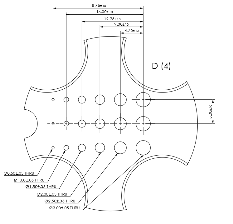
The general dimensions of the SPIRIT slice wedges are shown below in isolation. The SPIRIT Phantom contains features for determining slice thickness at the center of the phantom, referred to as the slice wedges. These features are designed in accordance with the NEMA MS 5-2018 standard [6].
Integrated Resolution Array
In addition, the slice wedges include a resolution array oriented orthogonally to the angled faces. This grid consists of 11 holes, linearly spaced, with diameters ranging from 3.00 mm ± 0.05 mm to 0.50 mm ± 0.05 mm.
Geometry
The slice wedges are positioned symmetrically at the centre of the phantom. Dimensional drawings (see figures) provide:
wedge angles and taper geometry,
reference coordinates relative to phantom datum surfaces,
resolution-grid hole placement and tolerances.
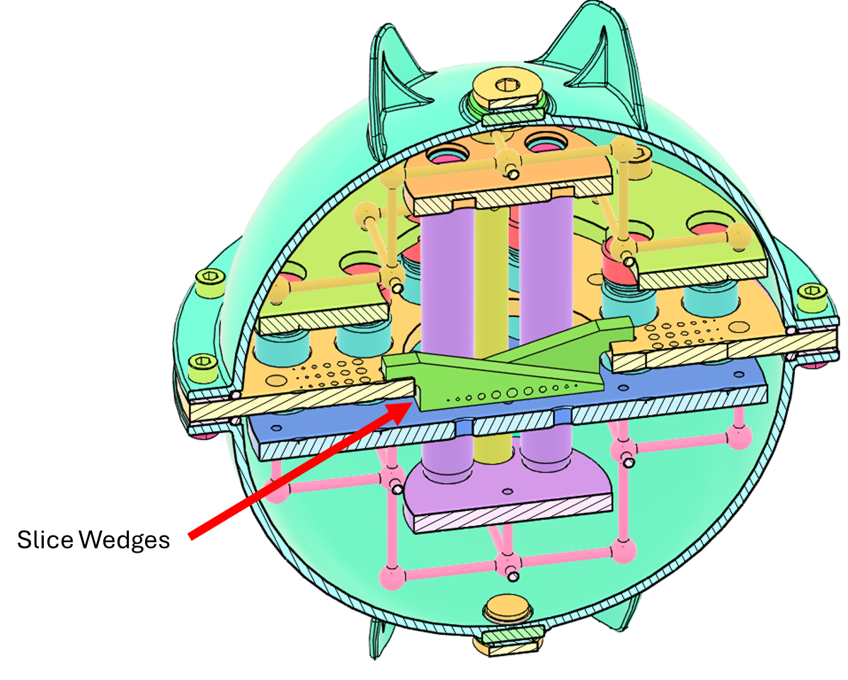
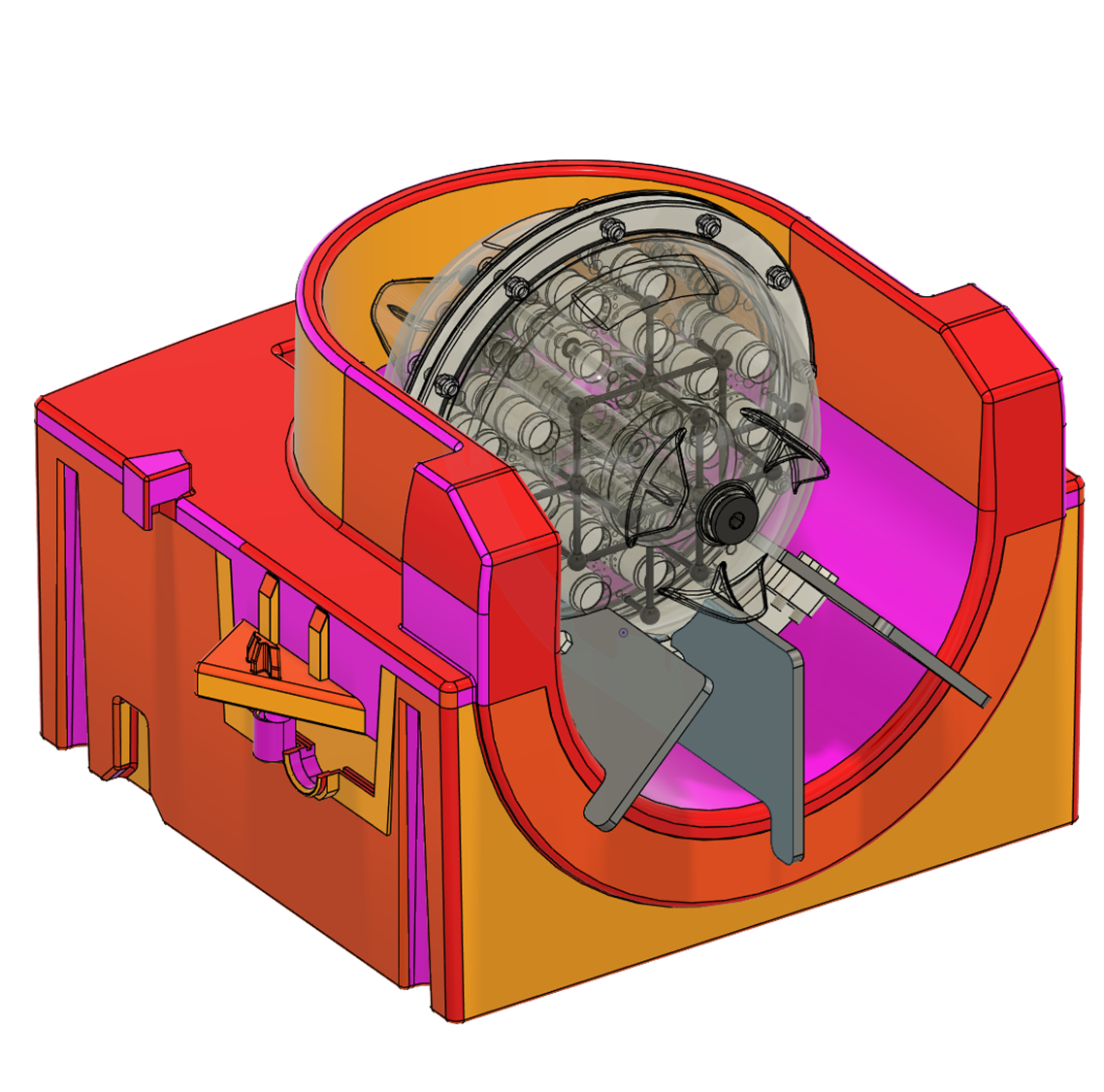
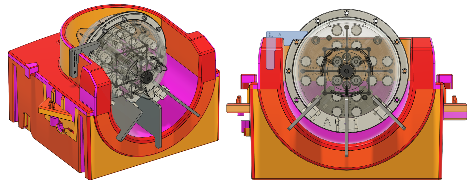
Section view of the SPIRIT phantom indicating the position of the slice wedges
The general dimensions of the SPIRIT slice wedges are shown below in isolation.
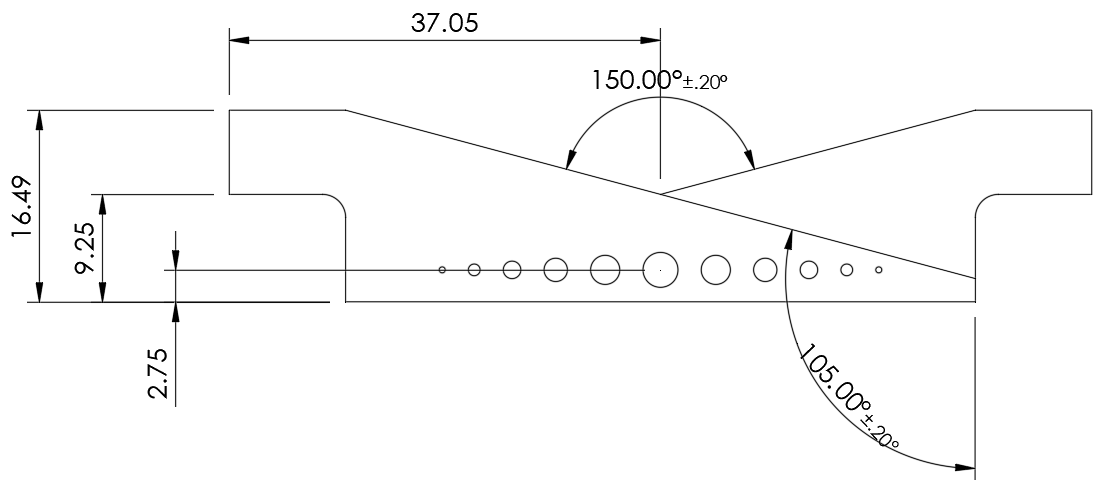
Dimensioned Drawing of the Slice Wedges indicating the angle of the slopes
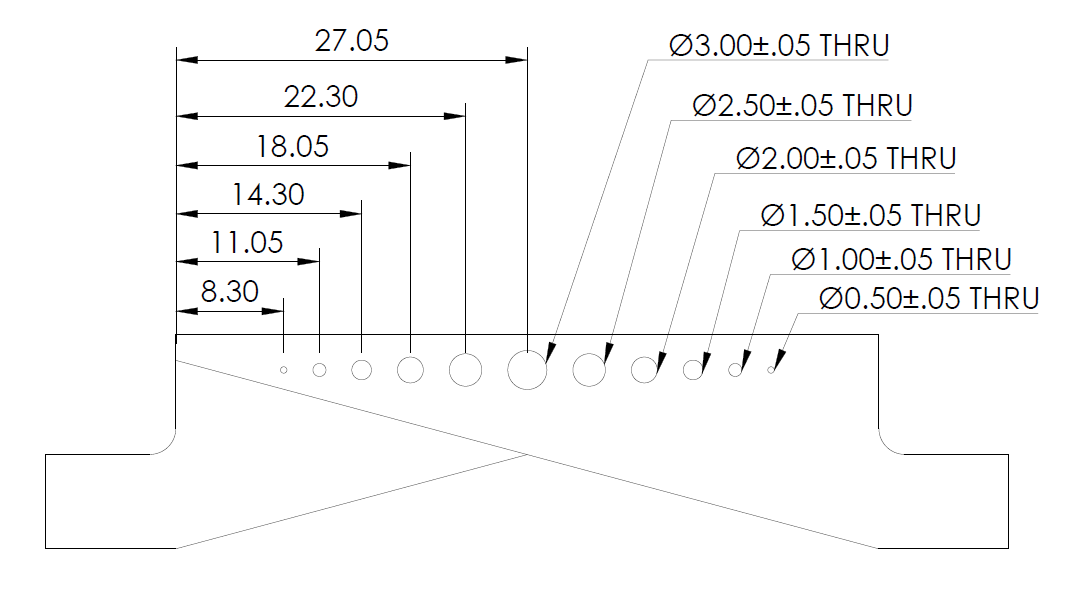
Dimensioned view showing the position and size of the slice wedge resolution array
About the Resolution Grids
Overview
Additional resolution-quantification features are located on the central plate of the SPIRIT Phantom. These allow assessment of image sharpness, partial-volume effects, system focus, and reconstruction quality across multiple directions.
Grid Layout
The central plate of the phantom incorporates a set of resolution grids arranged along the +X, -X, +Y, -Y axis.
Each grid consists of a 3 × 6 array of holes ranging from 3.00 mm ± 0.05 mm to 0.50 mm ± 0.05 mm, linearly spaced. The figures below illustrate their principal dimensions and layout.
Dimensional Features
Engineering drawings include:
grid origin and alignment relative to the phantom centre,
hole-to-hole spacing,
diameter tolerances,
plate thickness and depth profiles.
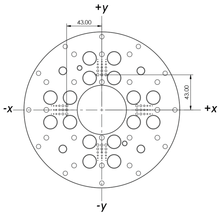
Zoomed-in isolated view of the grid showing hole spacing, diameters, and manufacturing tolerances.
About the Distortion Grid Fiducals
Overview
For geometric-distortion assessment, the SPIRIT Phantom contains a set of 52 fiducials arranged in a rectilinear 3D grid with a 34 mm center-to-center spacing.
This structure provides a precise framework for quantifying both in-plane and through-plane distortion in MR imaging systems.
Ball-and-Stick Assemblies - Upper and Lower Grids
The distortion grid consists of two identical ball-and-stick arrays (upper and lower), each containing 17 nodes.
The Grids include:
a solid 8 mm diameter sphere,
joined to adjacent nodes via 3 mm diameter rods.
When imaged using MRI, the nodes appear as signal voids, producing a high-contrast grid for geometric calibration.
Central-Plate Fiducials
The central plate includes eighteen (18) 6.00 mm diameter through-holes, which fill with phantom flood fill during use.
These appear as bright, high-signal features on MR images and provide cross-plate alignment markers.
Dimensional Features
Engineering drawings include:
3D grid layout,
node coordinates,
rod lengths and tolerances,
plate-through-hole positioning relative to phantom center.
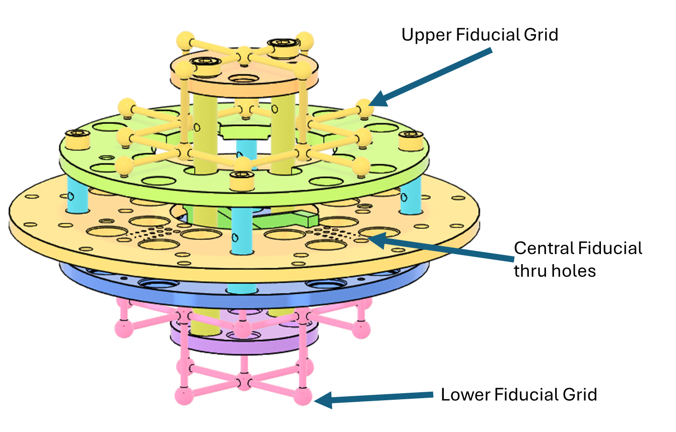
3D CAD representation of the SPIRIT Phantom showing the upper and lower fiducial grids along with the central-plate through-hole fiducials.
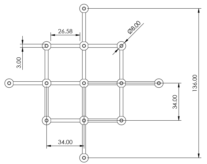
Technical drawing of the lower fiducial grid indicating key dimensions
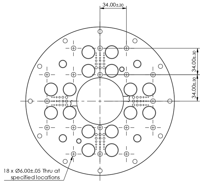
Excerpt of the SPIRIT Phantom central plate showing fiducial-hole positions.
Storage Instructions
Store In a dry and well-ventilated environment
Between 15 °C and 30 °C
Out of direct sunlight
If possible, store the phantom in the MRI magnet room. This will ensure that it is at thermal equilibrium with the room and provide the most reliable and reproducible results. Either store in the supplied box, or use the integrated legs to store on a flat surface such as a shelf.
Cleaning
If the SPIRIT phantom requires cleaning, they can be cleaned with soap, water, and a soft cloth or sponge to remove dirt and debris. Isopropanol may be used to remove sticky residue, however these may cause the labels to stop adhering to the surface.
Operating Instructions
Unpacking the equipment
The SPIRIT phantom is supplied in a protective cardboard packaging box. To unpack:
Inspect the outer packaging. If any damage is present, do not use the device. Please contact Gold Standard Phantoms.
Remove each component from the box.
Inspect each component for damage. If any damage is present, do not use the device. Please contact Gold Standard Phantoms.
Carefully remove the outer packaging. Do not use a sharp knife in case this cuts or punctures the device.
WARNING: The phantom weighs over 3 kg and will cause injury if dropped on bare feet or feet with open toed shoes. Take appropriate care when handling the phantom and wear suitable shoes that provide protection.
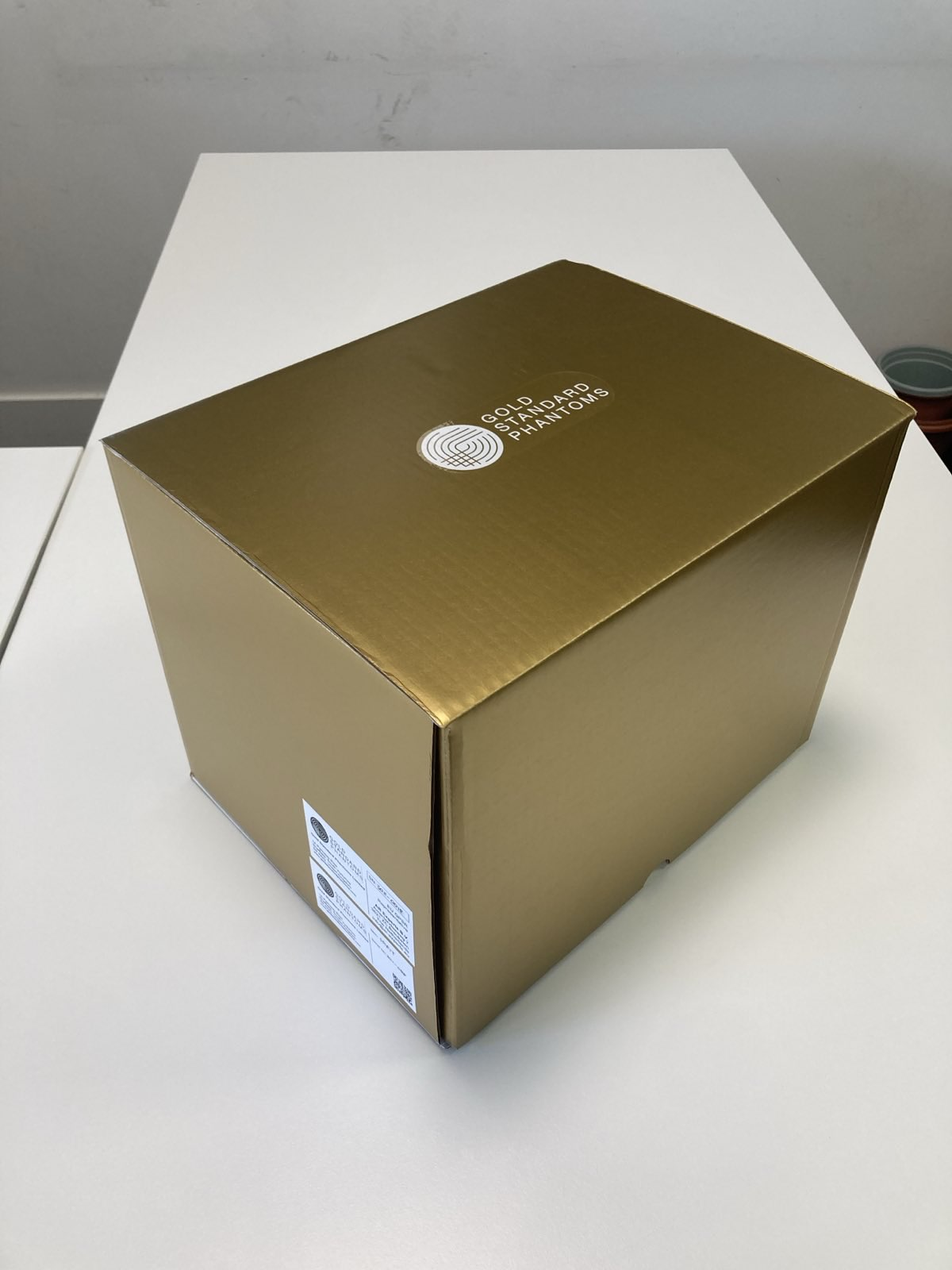
On receipt of the phantom remove the outer packaging to reveal the gold box
Opening the gold box reveals the following compenents
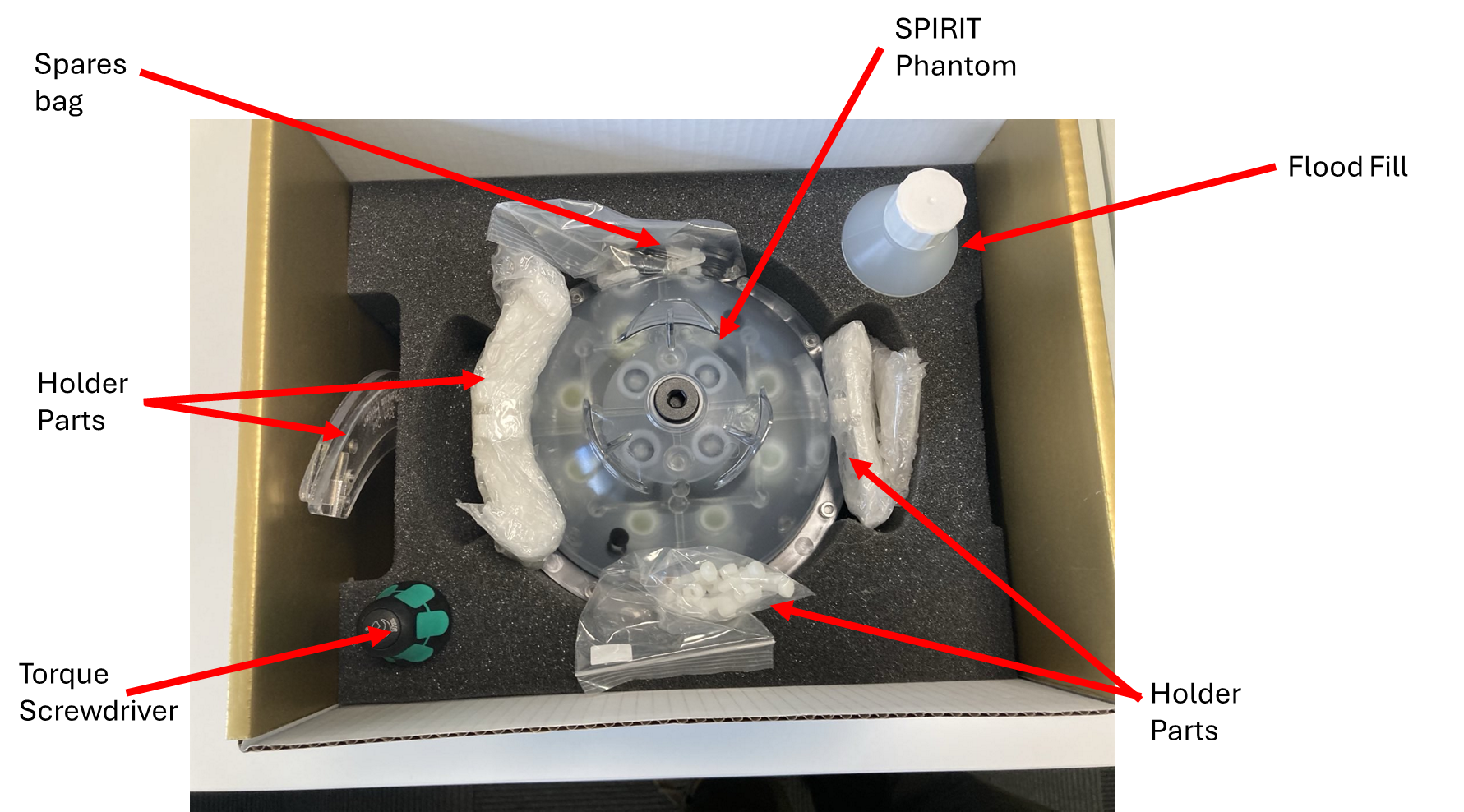
The SPIRIT box contains:
SPIRIT Phantom
Vial location key card
Paperwork including the SDS
Fixed Torque Screwdriver
Stainless Steel Hex Bits (6mm)
Spare Fixings
Phantom holder (to be assembled)
250 ml bottle of phantom flood fill liquid
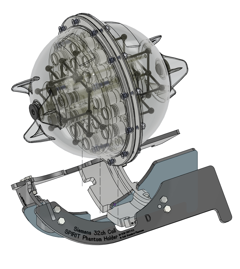
Holder Assembly and Use
The phantom holder requires assembly - instructions for this can be found at https://help.goldstandardphantoms.com/phantoms/spirit-phantom-holder-assembly-instructions
Phantom Use
This phantom is vendor-agnostic and is primarily designed for use in head coils.
For optimum performance, the phantom should be fitted into one of the coil specific Phantom holders with the SPIRIT product label facing up
Place the phantom onto the MRI scanner couch inside the head coil
Using the rotational alignment jig ensure the phantom is aligned in the coil
Align the MRI system’s laser with the middle of the phantom.
Move the phantom to magnet iso-centre.
Begin the MR examination.
When finished, store the phantom and holder.
Maintenance
Flood-fill top-up
The outer shell of the SPIRIT phantom is made from polycarbonate, which is permeable to water vapour. Over the course of several months water may evaporate from the flood-fill liquid that fills the phantom, resulting in bubbles forming. A bottle of flood-fill liquid is provided with the phantom, and can be used to top-up the phantom.
To top up the phantom:
Fit the supplied 6mm hex bit to the torque screwdriver.
Place the phantom on a flat surface.
Using the torque screwdriver, unscrew the cap (anticlockwise) from the filling port on the top of the phantom.
Add flood-fill liquid to the phantom until full. A pipette or syringe (not supplied) may aid.
Once full, re-fit the cap and tighten by hand a few turns.
Using the torque screwdriver, tighten the cap until the torque screwdriver “clicks”.
Check that there are no bubbles remaining in the phantom.
Repeat these steps as required until the phantom is bubble-free.
WARNING: The supplied torque screwdriver is pre-set to the correct tightening torque for the cap. Do not use any other tool to open or close the cap, damage to the phantom or cap may result.
Adverse Events
Any adverse events that occur during the use of this device should be reported both to Gold Standard Phantoms and the relevant Competent Authority of the member state.
Warranty
Standard Manufacturer’s Warranty (SMW)
The phantoms carry as 1 year manufacturer’s warranty. This covers assurance regarding quality / lifespan with a promise to repair or replace the SPIRIT under a standard RMA (return merchandise authorisation). Shipping and Duty charges may apply. This warranty shall be null and void in the event that the phantom sustains damage resulting from a fall or any other form of mishandling.
Disposal
Do not allow the contents of the phantom to enter drains, sewers, or watercourses. Dispose of this material and its container as hazardous waste. Disposal should be in accordance with local, state, or national legislation.
Citations
- ^ M. A. Boss et al., “Magnetic resonance imaging biomarker calibration service: proton spin relaxation times,” National Institute of Standards and Technology, May 2018. doi: 10.6028/nist.sp.250-97.
- ^ M. A. Boss et al., “Magnetic resonance imaging biomarker calibration service :,” National Institute of Standards and Technology (U.S.), Oct. 2022. doi: 10.6028/nist.sp.250-100.
- ^ A. L. Van Geet, “Calibration of the methanol and glycol nuclear magnetic resonance thermometers with a static thermistor probe,” Anal. Chem., vol. 40, no. 14, pp. 2227–2229, Dec. 1968, doi: 10.1021/ac50158a064.
- ^ D. S. Raiford, C. L. Fisk, and E. D. Becker, “Calibration of methanol and ethylene glycol nuclear magnetic resonance thermometers,” Anal. Chem., vol. 51, no. 12, pp. 2050–2051, Oct. 1979, doi: 10.1021/ac50048a040.
- ^ S. M. Sprinkhuizen, C. J. G. Bakker, and L. W. Bartels, “Absolute MR thermometry using time‐domain analysis of multi‐gradient‐echo magnitude images,” Magnetic Resonance in Med, vol. 64, no. 1, pp. 239–248, Jun. 2010, doi: 10.1002/mrm.22429.
- ^ National Electrical Manufacturers Association. (2018). Determination of slice thickness in diagnostic magnetic resonance imaging (NEMA MS 5-2018).